
Apoptosis and Caspases
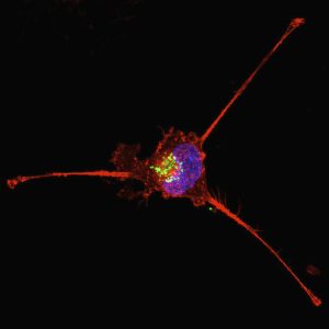
Apoptosis, also known as programmed cell death, is a highly regulated biological process that plays a critical role in maintaining normal tissue homeostasis and cellular balance. It is involved in several essential physiological functions, including immune system regulation, embryonic development, hormone-dependent tissue remodeling, metamorphosis, and the elimination of damaged or unwanted cells. In addition, apoptosis is activated in response to chemical stress, DNA damage, and various pathological conditions.
Defects in apoptotic signaling are strongly associated with many human diseases. Excessive apoptosis contributes to neurodegenerative disorders such as Alzheimer’s disease and Huntington’s disease, while insufficient apoptosis is linked to autoimmune diseases, ischemic injury, and multiple forms of cancer. Because of its major role in disease progression, apoptosis has become a central topic in molecular biology, cancer research, and therapeutic development.
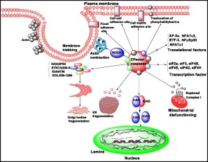
Apoptotic cell death is characterized by a series of well-defined biochemical and morphological changes. These include chromatin condensation, DNA fragmentation, reduction in cell volume, membrane blebbing, organelle compaction, and fragmentation of the nucleus. During the final stages of apoptosis, dying cells are recognized and removed by phagocytic cells without triggering inflammation. This non-inflammatory clearance distinguishes apoptosis from necrosis and is essential for maintaining tissue integrity.
The apoptotic process generally occurs in two major phases. The first phase involves cellular commitment to death following exposure to internal or external apoptotic signals. The second phase is the execution stage, during which specialized proteolytic enzymes systematically dismantle cellular structures. This execution mechanism is largely controlled by a family of cysteine proteases known as caspases.
Caspases and Their Biological Function
Caspases are a family of intracellular proteases that function as the primary executioners of apoptosis. The term “caspase” originates from “cysteine-dependent aspartate-specific protease,” reflecting both their catalytic mechanism and substrate specificity. These enzymes cleave target proteins after specific aspartic acid residues and are synthesized as inactive precursor molecules called procaspases.
Activation of procaspases occurs through proteolytic cleavage at conserved aspartate sites, generating active enzyme subunits that form functional protease complexes. Once activated, caspases initiate a proteolytic cascade that amplifies apoptotic signaling and leads to rapid cellular destruction.
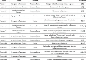
Phylogenetic and structural analyses classify caspases into several subfamilies, including:
- The inflammatory caspase group, such as caspase-1, caspase-4, and caspase-5
- The apoptotic executioner group, including caspase-3, caspase-6, and caspase-7
- The initiator caspases, such as caspase-8, caspase-9, and caspase-10
Among these enzymes, caspase-3 is considered one of the most important executioner caspases due to its ability to cleave multiple essential cellular proteins during apoptosis.
Caspase Activation Pathways
The understanding of caspase biology advanced significantly through studies on the nematode Caenorhabditis elegans. Researchers identified several genes involved in programmed cell death, including ced-3, ced-4, and ced-9. The ced-3 gene showed strong similarity to mammalian caspases, especially caspase-1, providing evidence that apoptotic pathways are evolutionarily conserved across species.
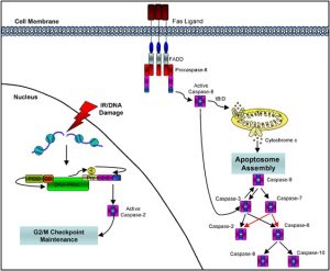
Apoptotic signaling can be triggered through two major pathways:
Extrinsic Pathway
The extrinsic apoptotic pathway begins with the activation of death receptors located on the cell membrane, such as CD95/Fas and tumor necrosis factor receptors (TNFR). Binding of specific ligands recruits adaptor proteins and activates initiator caspases, particularly caspase-8. Activated caspase-8 subsequently activates downstream executioner caspases, including caspase-3 and caspase-7.
Intrinsic Pathway
The intrinsic pathway is activated by intracellular stress signals such as DNA damage, oxidative stress, or mitochondrial dysfunction. This pathway involves mitochondrial release of pro-apoptotic factors that activate caspase-9, which then stimulates downstream executioner caspases.
Both pathways ultimately converge on a common execution phase involving widespread proteolytic cleavage of cellular substrates.
Structural Features of Caspases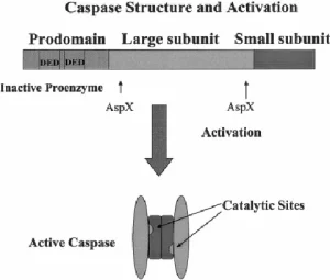
Caspases are synthesized as inactive zymogens composed of:
- An N-terminal prodomain
- A large catalytic subunit (p20)
- A small regulatory subunit (p10)
Following activation, cleavage removes inhibitory regions and allows formation of the active heterotetrameric enzyme complex. The catalytic activity of caspases depends on a highly conserved cysteine residue located within the active site.
Structural studies using X-ray crystallography revealed that active caspases contain substrate-binding pockets responsible for recognizing specific amino acid sequences. This specificity explains why different caspases target distinct intracellular proteins during apoptosis.
Caspase Substrates During Apoptosis
Activated caspases degrade a wide range of structural and regulatory proteins. Important substrates include:
- Poly(ADP-ribose) polymerase (PARP)
- DNA-dependent protein kinase (DNA-PK)
- Nuclear lamins
- Cytoskeletal proteins such as fodrin
- Retinoblastoma protein (Rb)
- Splicing-associated proteins like U1-70 kDa
Cleavage of these proteins contributes directly to the characteristic features of apoptosis, including nuclear fragmentation, chromatin condensation, DNA degradation, membrane blebbing, and cellular shrinkage.
For example, caspase-3-mediated cleavage of PARP inhibits DNA repair mechanisms, while caspase-6 targets nuclear lamins, leading to nuclear envelope breakdown.
Caspase Regulation and Inhibition
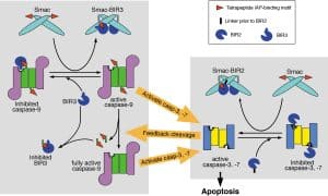
Because uncontrolled caspase activation can cause severe tissue damage, cells tightly regulate apoptotic pathways through several inhibitory mechanisms.
Viral Inhibitors
Certain viruses produce anti-apoptotic proteins such as CrmA and p35, which directly inhibit caspase activity. These viral proteins help infected cells avoid apoptosis, allowing viral replication to continue.
Bcl-2 Family Proteins
The Bcl-2 protein family regulates mitochondrial apoptosis by controlling caspase activation upstream. Anti-apoptotic members such as Bcl-2 and Bcl-xL suppress mitochondrial membrane permeabilization and inhibit downstream caspase activation.
Synthetic Caspase Inhibitors
Several peptide-based inhibitors, including Z-VAD-FMK and Ac-DEVD-CHO, are widely used in apoptosis research. These inhibitors block caspase activity and can prevent apoptotic cell death in experimental models. Caspase inhibitors are also being investigated for therapeutic applications in inflammatory diseases, liver injury, and neurodegenerative disorders.
Caspase Cascades and Hierarchical Activation
Caspases function through highly organized proteolytic cascades. Initiator caspases such as caspase-8 and caspase-9 are activated first in response to apoptotic signals. These enzymes subsequently activate executioner caspases including caspase-3, caspase-6, and caspase-7.
This hierarchical organization ensures rapid amplification of apoptotic signaling and coordinated cellular destruction. Different caspases may also activate one another through feedback amplification loops, increasing the efficiency of apoptosis.
Conclusion
Caspases are essential regulators and executioners of apoptosis, controlling the precise dismantling of cellular structures during programmed cell death. Their activation is fundamental for normal development, immune regulation, and tissue homeostasis. Dysregulation of caspase signaling contributes to a wide range of human diseases, including cancer, autoimmune disorders, and neurodegenerative conditions.
Advances in caspase biology have significantly improved the understanding of apoptotic mechanisms and opened new opportunities for targeted therapeutic strategies. Ongoing research continues to explore how selective modulation of caspase activity can be used in the treatment of cancer, inflammatory diseases, and degenerative disorders.