

Embryo selection remains one of the most important steps in assisted reproductive technology and in vitro fertilization (IVF). Improving embryo implantation rates while reducing the risk of multiple pregnancies is a major objective in reproductive medicine. Traditional embryo evaluation mainly relies on morphological assessment; however, this method has important limitations because embryo appearance alone cannot accurately predict developmental potential or chromosomal status. For this reason, current research is increasingly focused on identifying reliable noninvasive biomarkers through the analysis of spent embryo culture media.
Among the most promising biomarkers are microRNAs (miRNAs), which are small noncoding RNA molecules approximately 19–22 nucleotides in length. These molecules regulate gene expression at the post-transcriptional level and are involved in many essential cellular pathways, including differentiation, proliferation, metabolism, and apoptosis. miRNAs can be secreted into extracellular fluids inside exosomes, apoptotic bodies, or protein complexes that protect them from enzymatic degradation. Their remarkable stability in biological fluids such as plasma, serum, saliva, and culture media makes them attractive candidates for noninvasive diagnostic applications in reproductive biology.
Previous studies have demonstrated that miRNA expression is highly tissue-specific and plays a critical role in implantation, embryo development, decidualization, and endometrial receptivity. Altered miRNA expression profiles have also been associated with reproductive disorders including endometriosis, ectopic pregnancy, endometrial carcinoma, and preeclampsia. Human embryos and embryonic stem cells are known to express several miRNAs, and differences in miRNA expression have been reported between euploid and aneuploid blastocysts. Furthermore, secreted miRNAs detected in spent embryo culture media have been proposed as potential indicators of embryo viability, implantation competence, and chromosomal integrity.
Based on these observations, this study aimed to investigate the miRNA composition of spent culture media obtained from human euploid and aneuploid embryos using next-generation sequencing (NGS). The objective was to determine whether extracellular miRNA profiles could serve as noninvasive biomarkers for embryo selection in IVF programs.
Materials and Methods
Experimental Design
A total of 72 embryos obtained from 62 IVF patients were included in the study. Spent embryo culture media samples were collected and analyzed using two complementary experimental approaches. The investigation included:
- 36 spent media samples from day-3 euploid embryos
- 36 spent media samples from day-3 aneuploid embryos presenting more than three chromosomal abnormalities
- 36 matched control culture media samples without embryo exposure
For deep sequencing analysis, 48 pooled samples were processed by NGS:
- 16 euploid embryo media samples
- 16 aneuploid embryo media samples
- 16 control media samples without embryos
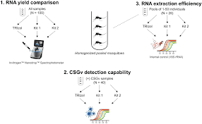
An additional 60 individual samples were evaluated using quantitative polymerase chain reaction (qPCR) for validation purposes. These included samples from euploid embryos, aneuploid embryos, and control media cultured in two different commercial embryo culture systems.
All procedures were approved by the institutional ethics committee, and informed consent was obtained from all participating patients.
Embryo Culture and Blastomere Biopsy
Following oocyte retrieval, fertilization was performed using either conventional IVF or intracytoplasmic sperm injection (ICSI), depending on sperm quality parameters. Embryo development was monitored daily, and embryos were cultured until day 3 using commercially available embryo culture media supplemented with human serum albumin.
Embryo quality assessment included evaluation of:
- Blastomere number
- Fragmentation percentage
- Blastomere symmetry
- Multinucleation
- Cytoplasmic morphology
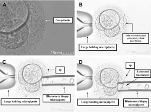
Only embryos containing more than six nucleated blastomeres and less than 20% fragmentation were selected for biopsy. A single blastomere was removed using laser-assisted zona pellucida opening techniques. After biopsy, embryos continued to develop in culture until day 5 prior to embryo transfer.
Fluorescence In Situ Hybridization (FISH) Analysis
Preimplantation genetic screening was performed using fluorescence in situ hybridization (FISH) to analyze chromosomes 13, 15, 16, 17, 18, 21, 22, X, and Y. Embryos were classified as:
- Euploid (normal): Correct chromosomal signal pattern
- Aneuploid (abnormal): Presence of abnormal chromosome numbers
Microscopic analysis was conducted using fluorescence imaging systems equipped with multiple filter configurations for chromosome visualization.
Collection of Embryo-Conditioned Culture Media
Spent embryo culture media were collected after embryo biopsy. Approximately 50 µL of media from each embryo culture droplet was transferred into sterile tubes. Samples were centrifuged to eliminate cellular contamination, and supernatants were stored at −80°C until molecular analysis.
Matched control media that had not been exposed to embryos underwent the same processing protocol.
Next-Generation Sequencing (NGS)
Small RNA libraries were prepared according to Ion Torrent sequencing protocols. Equal quantities of pooled samples from euploid embryos, aneuploid embryos, and control media were combined to create an equimolar sequencing library.
Sequencing reads underwent several preprocessing steps:
- Adapter trimming
- Removal of sequencing artifacts
- Selection of small RNA fragments between 17 and 30 nucleotides
- Alignment against the miRBase database
miRNA identification and quantification were performed using the miRDeep2 bioinformatics platform.
RNA Extraction and qPCR Validation
miRNAs were isolated from spent culture media using the QIAGEN miRNeasy extraction kit. Reverse transcription and complementary DNA amplification were subsequently performed.
Quantitative real-time PCR (RT-PCR) analysis focused on two highly abundant miRNAs identified during NGS screening:
- miR-181b-5p
- miR-191-5p
Expression levels were normalized using the housekeeping control SNORD96, and relative expression changes were calculated using the 2^-ΔΔCt method.
Additional amplification experiments confirmed the linearity and reproducibility of the qPCR assays.
Bioinformatics and Functional Analysis
Identified miRNAs were analyzed using several bioinformatics resources, including:
- miRBase
- miRTarBase
- GeneMANIA
Potential target genes and biological pathways associated with miR-181b-5p and miR-191-5p were investigated. Functional enrichment analysis identified pathways related to:
- Embryo development
- Cell cycle regulation
- Apoptosis
- Mitochondrial organization
- Stress response signaling
Statistical analyses were conducted using SPSS software.
Results
Identification of miRNAs in Spent Embryo Culture Media
NGS analysis identified 53 known human miRNAs across all study groups. Among these, miR-181b-5p and miR-191-5p represented nearly 69% of all detected sequencing reads, making them the most abundant extracellular miRNAs identified in the culture media.
Although direct read counts initially suggested slightly higher abundance in euploid embryos, normalization analysis revealed no statistically significant differences between:
- Euploid embryo media
- Aneuploid embryo media
- Control media without embryos
Most detected miRNAs exhibited expression levels close to background noise.
qPCR Validation Findings
Individual qPCR validation experiments confirmed that miR-181b-5p and miR-191-5p were present at similar levels in both embryo-conditioned and control media samples.
Importantly, comparable miRNA expression profiles were observed regardless of:
- Embryo chromosomal status
- Culture media formulation
- Presence or absence of embryos
These findings strongly suggested that the detected miRNAs originated primarily from the commercial culture media itself rather than from embryo secretion.
Additional fractionation experiments demonstrated that these miRNAs were associated with high-molecular-weight protein complexes, indicating that they were protein-bound rather than freely circulating molecules.
Biological Significance of miR-181b-5p and miR-191-5p
Functional target analysis revealed that miR-181b-5p and miR-191-5p potentially regulate genes involved in critical developmental and cellular processes.
Important target genes included:
- MDM4 : apoptosis regulation
- SOX4 : embryonic development
- CDK6 : cell cycle progression
- SATB1 : chromatin organization
- BCL2 and MCL1 mitochondrial and apoptotic pathways
Gene enrichment analysis demonstrated strong associations with:
- Embryonic development
- Mitochondrial membrane organization
- Cellular signaling pathways
- Protein transport
- Stress response mechanisms
These findings suggest that even externally derived miRNAs present in embryo culture media may potentially influence embryo physiology and developmental programming.
Discussion
The use of spent embryo culture media as a noninvasive source of biomarkers has generated considerable interest in IVF research. However, the present study demonstrated that the majority of detectable extracellular miRNAs in embryo culture media are likely derived from the commercial media formulation itself rather than secreted by developing embryos.
No significant differences were identified between euploid and aneuploid embryos regarding miRNA abundance, limiting the utility of these molecules as reliable biomarkers for embryo chromosomal screening.
Interestingly, several miRNAs were already present in unused culture media before embryo exposure, reinforcing the possibility of external contamination or media-derived molecular background signals. This finding raises important concerns regarding the interpretation of molecular analyses performed on spent embryo culture systems.
The study also highlights the possibility that commercially available embryo culture media may contain biologically active molecules capable of influencing embryo gene expression and developmental pathways. Previous research has shown that maternal extracellular miRNAs can regulate embryo transcriptomic activity and implantation processes, supporting the concept that environmental miRNAs may alter embryonic physiology.
Although the study was limited by sample size, the results emphasize the need for greater transparency regarding the molecular composition of commercial embryo culture media used in IVF laboratories.
Conclusion
Using next-generation sequencing and RT-PCR analysis, this study demonstrated that extracellular miRNAs detected in spent human embryo culture media are predominantly derived from commercial culture media rather than secreted by embryos themselves. No significant differences in miRNA abundance were observed between euploid and aneuploid embryos.
These findings suggest that miRNAs currently have limited diagnostic value as noninvasive biomarkers for embryo chromosomal assessment in IVF. The study also raises important concerns about the unknown molecular composition of embryo culture media and its possible influence on embryo development and implantation outcomes.