
Introduction to Central Nervous System Infections
Acute infections affecting the central nervous system (CNS) are considered major medical emergencies because delayed diagnosis or treatment can lead to severe neurological damage, permanent disability, or death. CNS infections include a broad group of disorders such as meningitis, encephalitis, and brain abscesses. These conditions can rapidly compromise brain function and significantly reduce patient survival and quality of life.
The central nervous system consists of the brain, spinal cord, optic nerves, and their surrounding protective membranes. These structures are enclosed within the skull and vertebral column, providing mechanical protection against external injury. The brain itself contains two major regions: the cerebrum and cerebellum, while the spinal cord serves as the principal communication pathway between the brain and peripheral nervous system.
Three protective connective tissue layers known as the meninges surround the CNS. The innermost layer, called the pia mater, lies directly on the surface of the brain and spinal cord. The middle layer is the arachnoid mater, while the outermost and strongest layer is the dura mater. Between these membranes are several anatomical spaces that play important roles in infection spread and cerebrospinal fluid circulation.
The subarachnoid space, located between the pia and arachnoid membranes, contains cerebrospinal fluid (CSF) and extends continuously around the entire CNS. The subdural space lies between the arachnoid and dura mater, whereas the epidural space is positioned between the dura and the skull. Certain infectious microorganisms can invade these spaces directly or through bloodstream dissemination, resulting in meningitis, abscess formation, or inflammatory complications.
Cerebrospinal Fluid and Blood–Brain Barrier Function
Cerebrospinal fluid is continuously produced by the choroid plexus inside the ventricular system of the brain. The CSF circulates through the lateral ventricles, third ventricle, and fourth ventricle before entering the subarachnoid space, where it surrounds and protects the brain and spinal cord. Eventually, CSF is reabsorbed into the bloodstream through arachnoid villi located within the dural venous sinuses.
A critical protective structure in CNS physiology is the blood–brain barrier (BBB). This barrier is formed by specialized endothelial cells joined by tight junctions that strictly regulate molecular transport between the bloodstream and neural tissues. The BBB limits the entry of pathogens, toxins, and many pharmacological agents into the brain while maintaining neural homeostasis.
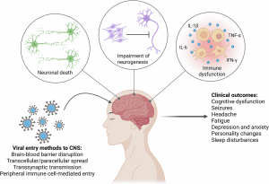
CNS infections develop when microorganisms overcome or disrupt the blood–brain barrier. This may occur through inflammatory cytokine release, endothelial damage, microvascular obstruction, tissue necrosis, or direct microbial interactions with endothelial receptors. Certain pathogens, including Escherichia coli, mycobacteria, and spirochetes, possess specialized mechanisms that facilitate penetration across the BBB.
Understanding blood–brain barrier physiology is essential because effective antimicrobial therapy for CNS infections requires antibiotics capable of achieving therapeutic concentrations within the CNS.
Routes of CNS Infection
Central nervous system infections generally occur through hematogenous dissemination, where bacteria or viruses spread via the bloodstream from distant infection sites. Primary infections involving the respiratory tract, lungs, middle ear, mastoid sinuses, gastrointestinal tract, kidneys, or skin may seed microorganisms into the CNS circulation.
In children, sinusitis and middle ear infections are among the most common predisposing conditions leading to transient bacteremia and CNS invasion. Infections affecting the paranasal sinuses or mastoid regions can also spread through thrombophlebitis involving cortical veins and dural venous sinuses.
Microorganisms may enter the CNS through several additional pathways:
- Direct extension from nearby infections
- Skull base fractures causing CSF leakage
- Neurosurgical procedures
- Colonized medical devices such as shunts or drainage systems
- Congenital malformations including spina bifida or dermal sinus tracts
- Intraneural spread, as observed with rabies virus, herpes simplex virus, and polioviruses
Brain abscesses may develop either through direct extension of infection or through septic emboli associated with endocarditis or pulmonary abscesses.
Acute Bacterial Meningitis
Definition and Clinical Importance
Acute bacterial meningitis is a severe inflammatory infection involving the pia mater, arachnoid membranes, cerebrospinal fluid, and surrounding CNS tissues. The inflammatory response is usually pyogenic, characterized by massive neutrophilic infiltration and purulent exudate formation.
The disease typically progresses rapidly and presents with:
- Fever
- Severe headache
- Neck stiffness
- Vomiting
- Photophobia
- Altered mental status
- Neurological deficits
Without prompt antimicrobial treatment, bacterial meningitis is almost universally fatal.
Epidemiology of Bacterial Meningitis
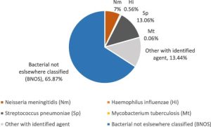
Several bacterial pathogens are responsible for most cases of acute bacterial meningitis.
Streptococcus pneumoniae
Streptococcus pneumoniae is the leading cause of community-acquired bacterial meningitis in many countries. It affects nearly all age groups except neonates and is strongly associated with:
- Alcoholism
- Diabetes mellitus
- HIV infection
- Chronic kidney disease
- Splenectomy
- Cerebrospinal fluid leaks
- Immunosuppression
Transmission occurs primarily through respiratory droplets and nasopharyngeal colonization.
Neisseria meningitidis
Neisseria meningitidis colonizes the nasopharynx and spreads through respiratory secretions. Humans are the only reservoir.
High-risk populations include:
- Military recruits
- College students in dormitories
- Household contacts
- Complement-deficient individuals
- Travelers to endemic regions
Meningococcal disease may present as:
- Meningitis
- Septicemia
- Pneumonia
- Fulminant shock
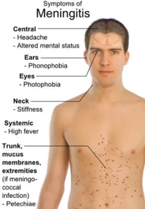
Characteristic petechial or purpuric skin rashes may appear during meningococcemia.
Haemophilus influenzae Type b
Haemophilus influenzae type b was historically a major cause of childhood meningitis before widespread vaccination programs dramatically reduced disease incidence.
Risk factors include:
- Immunodeficiency
- HIV infection
- Chronic renal disease
- Malignancy
- Sickle cell disease
Vaccination against Hib remains one of the most successful preventive strategies in infectious disease medicine.
Listeria monocytogenes
Listeria monocytogenes commonly affects:
- Neonates
- Elderly individuals
- Pregnant women
- Immunocompromised patients
- Organ transplant recipients
Transmission is usually foodborne and associated with contaminated dairy products, processed meats, and unpasteurized foods.
Other Important Pathogens
Additional bacterial causes include:
- Escherichia coli
- Streptococcus agalactiae
- Klebsiella
- Pseudomonas
- Staphylococcus aureus
These organisms are particularly important in neonates, hospitalized patients, and individuals undergoing neurosurgical procedures.
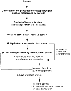
Pathogenesis of Bacterial Meningitis
The development of meningitis generally follows several sequential stages:
- Nasopharyngeal colonization
- Bloodstream invasion
- Survival in circulation
- Blood–brain barrier penetration
- Entry into the subarachnoid space
- Uncontrolled bacterial replication
- Intense inflammatory response
Bacterial virulence factors such as capsules, pili, fimbriae, IgA proteases, and outer membrane proteins play essential roles in host invasion and immune evasion.
Once bacteria enter the CSF, immune defenses become limited because cerebrospinal fluid contains relatively low concentrations of antibodies, complement proteins, and phagocytic cells.
Bacterial components such as lipopolysaccharide (LPS), peptidoglycan, and teichoic acid trigger massive cytokine production, particularly:
- Tumor necrosis factor-alpha (TNF-α)
- Interleukin-1 (IL-1)
These inflammatory mediators increase blood vessel permeability, recruit neutrophils, and promote cerebral edema, vasculitis, ischemia, and tissue necrosis.
Clinical Manifestations
The classic triad of bacterial meningitis includes:
- Fever
- Neck stiffness
- Altered mental status
Additional manifestations may include:
- Severe headache
- Photophobia
- Nausea and vomiting
- Seizures
- Focal neurological deficits
- Coma
Presentation varies depending on age and pathogen. Neonates and elderly patients often show atypical or nonspecific symptoms such as irritability, confusion, lethargy, or poor feeding.
Meningococcal septicemia may progress rapidly to:
- Disseminated intravascular coagulation (DIC)
- Shock
- Purpura fulminans
- Waterhouse–Friderichsen syndrome
Diagnosis of CNS Infections
Lumbar Puncture and CSF Analysis
Lumbar puncture remains the cornerstone of meningitis diagnosis.
Typical bacterial meningitis CSF findings include:
- Elevated opening pressure
- High neutrophil count
- Increased protein concentration
- Low glucose levels
- Turbid or purulent appearance
Gram stain and bacterial culture are essential for pathogen identification.
Normal CSF contains very few leukocytes and low protein levels. In bacterial meningitis, white blood cell counts may exceed:
10,000 cells/mm3
with neutrophilic predominance.
CSF glucose commonly falls below:
40 mg/dL
due to impaired glucose transport and microbial metabolism.
Neuroimaging
Computed tomography (CT) and magnetic resonance imaging (MRI) are primarily used to evaluate complications such as:
- Brain abscess
- Cerebral infarction
- Hydrocephalus
- Subdural empyema
- Cerebral edema
- Herniation risk
MRI is particularly sensitive for detecting cerebritis and ischemic lesions.
Treatment of Acute Bacterial Meningitis
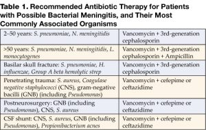
Immediate empirical antimicrobial therapy is essential and should never be delayed when bacterial meningitis is strongly suspected.
Third-generation cephalosporins such as:
- Ceftriaxone
- Cefotaxime
are commonly used because they penetrate inflamed meninges effectively.
Vancomycin is frequently added to cover penicillin-resistant pneumococci. Ampicillin is included when Listeria monocytogenes is suspected.
Treatment selection depends on:
- Patient age
- Immune status
- Healthcare-associated exposure
- Local antimicrobial resistance patterns
Adjunctive dexamethasone therapy may reduce neurological complications and hearing loss in selected patients, particularly children with Hib meningitis.
Complications of CNS Infections
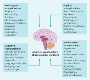
Serious complications include:
- Elevated intracranial pressure
- Cerebral edema
- Hydrocephalus
- Vasculitis
- Cerebral infarction
- Seizures
- Brain herniation
- Permanent neurological impairment
Raised intracranial pressure can rapidly become fatal if untreated.
Management strategies include:
- Elevation of the head
- Mechanical ventilation
- Hyperventilation
- Mannitol administration
- Hypertonic saline therapy
- Intracranial pressure monitoring
Conclusion
Central nervous system infections remain among the most severe infectious diseases in clinical medicine. Acute bacterial meningitis, encephalitis, and intracranial abscesses require rapid recognition, immediate diagnostic evaluation, and aggressive antimicrobial therapy. Understanding CNS anatomy, blood–brain barrier physiology, microbial pathogenesis, and inflammatory mechanisms is essential for accurate diagnosis and effective treatment.
Despite major advances in vaccination, antimicrobial therapy, neuroimaging, and intensive care, CNS infections continue to cause substantial morbidity and mortality worldwide. Early diagnosis and prompt treatment remain the most important factors for improving patient outcomes.