
Introduction to Hantaviruses
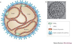
Orthohantavirus are enveloped RNA viruses that belong to the International Committee on Taxonomy of Viruses order Bunyavirales and the Hantaviridae family. These viruses possess a negative-sense, single-stranded RNA genome divided into three segments: large (L), medium (M), and small (S). The L segment encodes the viral RNA-dependent RNA polymerase, while the M segment produces a glycoprotein precursor that is later processed into the Gn and Gc envelope glycoproteins. The S segment encodes the nucleoprotein (NP) and, in some species, a non-structural protein known as NSs.
Hantavirus particles are pleomorphic, enveloped virions measuring approximately 70–160 nm in diameter. The viral surface contains tetrameric spike complexes composed of Gn and Gc glycoproteins, which are essential for host-cell attachment, viral entry, and assembly.
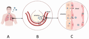
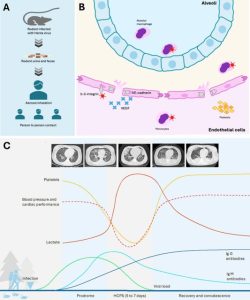
Rodents are the primary natural reservoirs of hantaviruses, and each viral species generally demonstrates a strong association with a specific rodent host. Environmental disturbances, climate variation, and ecological changes can significantly influence rodent population dynamics and consequently increase the probability of hantavirus outbreaks. Human infection usually occurs through inhalation of aerosolized particles contaminated with rodent urine, saliva, or feces. Humans are commonly considered dead-end hosts, although person-to-person transmission has been documented for Andes orthohantavirus infections.
Hantavirus Diseases in Humans
Hantaviruses are responsible for two major zoonotic diseases in humans:
- Hemorrhagic Fever with Renal Syndrome (HFRS)
- Hantavirus Cardiopulmonary Syndrome (HCPS)
Globally, approximately 200,000 hantavirus infections are reported every year. More than 50 hantavirus species have been identified, with over 24 recognized as pathogenic to humans.
Hemorrhagic Fever with Renal Syndrome (HFRS)
HFRS is mainly associated with Eurasian hantaviruses such as:
- Hantaan orthohantavirus
- Dobrava–Belgrade orthohantavirus
- Puumala orthohantavirus
Case fatality rates range from 0.1% to 15%, depending on the viral strain and disease severity. Clinical manifestations typically progress through febrile, hypotensive, oliguric, polyuric, and convalescent phases. Severe cases involve acute kidney injury, proteinuria, hematuria, thrombocytopenia, and vascular leakage.
Hantavirus Cardiopulmonary Syndrome (HCPS)
HCPS occurs predominantly in the Americas and is primarily caused by:
- Sin Nombre orthohantavirus
- Andes orthohantavirus
HCPS is characterized by high mortality rates reaching 20–40%. Patients initially develop influenza-like symptoms followed by severe respiratory distress, pulmonary edema, hypotension, cardiogenic shock, and respiratory failure.
Unlike HFRS, HCPS progresses rapidly and often results in life-threatening pulmonary complications. Survivors may continue experiencing respiratory dysfunction years after recovery.
Pathogenesis and Endothelial Dysfunction
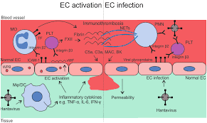
Microvascular endothelial cells are the primary targets of hantavirus infection. Although the viruses do not directly destroy infected endothelial cells, they trigger profound vascular dysfunction and increased capillary permeability.
One major pathogenic mechanism involves dysregulation of vascular endothelial growth factor A (VEGF-A), which contributes to endothelial leakage and pulmonary edema. Elevated levels of VEGF and inflammatory mediators have been linked with severe disease progression.
Hantavirus infections are also associated with:
- Thrombocytopenia
- Leukocyte activation
- Cytokine overproduction
- Increased vascular permeability
- Coagulation abnormalities
These alterations contribute to systemic vascular leakage, tissue edema, and organ dysfunction.
Cytokine Storm and Inflammatory Response
A hallmark of severe hantavirus disease is the development of a strong proinflammatory immune response known as a cytokine storm. Elevated concentrations of inflammatory mediators are detected in serum, tissues, and organs of infected individuals.
Important cytokines and inflammatory markers include:
- IL-1β
- IL-2
- IL-6
- IL-8
- TNF
- IFN-γ
- CXCL9
- CXCL10
- Nitric oxide
- C-reactive protein (CRP)
High IL-6 levels are strongly associated with severe HFRS and fatal HCPS outcomes. Excessive cytokine production promotes immune-cell recruitment, endothelial damage, platelet migration, and vascular leakage.
HCPS patients generally display a more intense innate immune activation and stronger Th1-associated cytokine profile compared with mild HFRS cases.
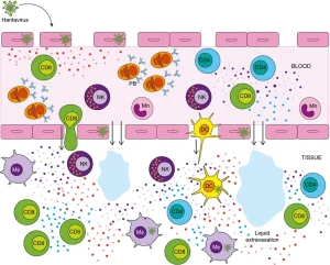
Innate Immune Response to Hantavirus Infection
Recognition of Hantaviruses by Innate Immunity
Host innate immune receptors detect hantavirus infection through recognition of viral RNA structures. Several pattern-recognition receptors
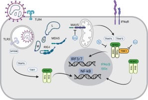
participate in antiviral defense, including:
- Toll-like receptor 3 (TLR3)
- Retinoic acid-inducible gene I (RIG-I)
- Melanoma differentiation-associated protein 5 (MDA5)
- Protein kinase R (PKR)
These receptors activate signaling pathways leading to type I interferon production and expression of interferon-stimulated genes (ISGs).
However, pathogenic hantaviruses have evolved sophisticated immune evasion mechanisms. For example:
- ANDV nucleoprotein interferes with interferon regulatory signaling
- NSs proteins inhibit MAVS activation
- Viral Gn cytoplasmic tails suppress interferon pathways
- Viral proteins inhibit PKR activation and stress granule formation
These mechanisms delay antiviral responses and promote viral replication during early infection.
Natural Killer (NK) Cells
Natural killer cells are essential innate lymphocytes involved in early antiviral defense. During hantavirus infection, NK cells become highly activated and produce:
- Granzyme B
- Perforin
- IFN-γ
- TNF
In patients infected with PUUV, activated CD56dim NK cells display increased expression of cytotoxicity markers such as CD69, NKp30, and NKp46.
Despite this strong activation, infected endothelial cells often resist NK-mediated apoptosis due to viral inhibition of TRAIL signaling and caspase activation pathways.
Dendritic Cells and Macrophages
Hantaviruses infect dendritic cells (DCs) and macrophages, promoting their maturation and inflammatory activation.
Infected dendritic cells show increased expression of:
- MHC class I and II molecules
- Costimulatory molecules
- Adhesion molecules
These activated antigen-presenting cells produce inflammatory cytokines and improve antigen cross-presentation, facilitating CD8+ T-cell activation.
Pathogenic hantaviruses replicate efficiently in monocytes and dendritic cells, unlike low-pathogenic strains such as Prospect Hill orthohantavirus. This replication capacity appears linked to viral pathogenicity.
Macrophages may also contribute to ANDV transmission because viral antigens have been detected in alveolar macrophages and salivary gland tissues.
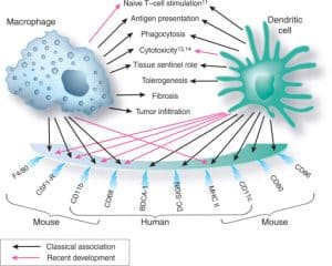
Adaptive Immune Response During Hantavirus Infection
CD8+ T-Cell Response
Cytotoxic CD8+ T lymphocytes play a major role in antiviral immunity during hantavirus infection. Strong virus-specific CD8+ T-cell responses are associated with:
- Granzyme B secretion
- Perforin release
- Viral clearance
- Long-lasting immune memory
NP-specific CD8+ T cells may persist for more than 15 years after infection, indicating durable cellular immunity.
Although these cells contribute to viral elimination, excessive cytotoxic activity may also participate in endothelial injury and vascular leakage during severe disease.
Animal studies suggest that CD8+ T-cell responses are important for protection against hantavirus infection, especially in cross-protective immunity between different hantavirus species.
CD4+ T-Cell Response
CD4+ T-helper cells contribute to immune regulation during hantavirus disease. Mixed Th1/Th2 cytokine profiles are commonly observed in infected individuals.
Research also indicates an important role for regulatory T cells (Tregs) in controlling immunopathology. Increased expression of FOXP3 and immune checkpoint molecules such as PD-1 and CTLA-4 suggests that immune suppressive mechanisms attempt to limit excessive inflammation.
Differences in Treg activation may partly explain variations in disease severity between PUUV and DOBV infections.
B-Cell and Antibody Responses
Hantavirus infection induces a strong humoral immune response. IgM antibodies appear early after symptom onset, followed by long-lasting IgG production.
The initial antibody response mainly targets the viral nucleoprotein, while neutralizing antibodies against Gn and Gc glycoproteins are associated with protective immunity and survival.
High neutralizing antibody titres correlate with:
- Better clinical outcomes
- Increased survival rates
- Long-term protection
Interestingly, survivors of ANDV infection may continue showing increasing neutralizing antibody levels years after disease recovery.
Differences in IgG subclass responses have also been observed:
- PUUV infections are dominated by IgG1
- HCPS caused by SNV is associated mainly with IgG3
Massive plasmablast expansion has been reported during acute infection, along with the appearance of autoantibodies and non-virus-specific antibody responses.
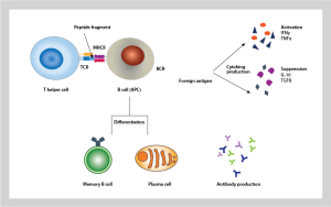
Current Therapeutic Strategies
Currently, no FDA-approved antiviral therapy exists for hantavirus diseases. Clinical management mainly relies on:
- Supportive care
- Hemodynamic stabilization
- Oxygen therapy
- Extracorporeal membrane oxygenation (ECMO) in severe HCPS
Passive immunotherapy using convalescent plasma rich in neutralizing antibodies has shown promising reductions in case fatality rates.
Monoclonal antibodies targeting ANDV glycoproteins have demonstrated strong protective effects in experimental animal models. Polyclonal human IgG generated through DNA-vaccinated transchromosomal bovines has also shown significant neutralizing activity.
Because cytokine storms contribute heavily to disease severity, immunomodulatory therapies targeting IL-6 and inflammatory pathways are currently considered promising future treatment approaches.
Vaccine Development Against Hantaviruses
Challenges in Vaccine Development
Despite extensive research, no universally approved hantavirus vaccine exists. Major challenges include:
- Limited animal models
- Incomplete understanding of immunopathology
- High biosafety requirements
- Diversity among hantavirus species
Experimental Vaccine Strategies
Several vaccine technologies are currently under investigation:
Inactivated Vaccines
- Hantavax
- Bivalent HTNV/SEOV vaccines
Recombinant Protein Vaccines
- NP-based vaccines
- Glycoprotein subunit vaccines
Viral Vector Vaccines
- Adenovirus vectors
- Vesicular stomatitis virus (VSV) vectors
Virus-Like Particle Vaccines
- VLP-based immunization platforms
DNA Vaccines
DNA vaccines targeting HTNV, PUUV, SNV, and ANDV have shown promising immunogenicity and protective responses in animal models and clinical trials.
Importance of Cellular Immunity in Vaccination
Neutralizing antibodies are essential for protection, but increasing evidence demonstrates that cellular immunity is equally important.
Several vaccine studies have shown protection even in the absence of detectable neutralizing antibodies, indicating a crucial role for:
- CD8+ cytotoxic T cells
- IFN-γ secretion
- TNF-mediated antiviral activity
- Memory T-cell responses
Cross-protective immunity between different hantavirus species also appears strongly linked to cellular immune mechanisms.
These observations support the possibility of developing a future pan-hantavirus vaccine capable of protecting against multiple pathogenic species.
Conclusion
Hantavirus diseases represent severe emerging zoonotic infections characterized by vascular leakage, cytokine storms, and dysregulated immune responses. Both innate and adaptive immunity participate in viral control and disease pathogenesis.
While neutralizing antibodies are strongly associated with survival and long-term protection, cytotoxic T cells and inflammatory mediators also play fundamental roles in viral clearance and immunopathology.
Recent advances in monoclonal antibodies, DNA vaccines, viral vector vaccines, and immunotherapeutic strategies provide promising opportunities for future prevention and treatment. However, a deeper understanding of protective immunity, immune regulation, and cross-reactive responses remains essential for the successful development of safe and effective hantavirus vaccines and therapies.