
SARS-CoV-2 and Coronavirus Spike Proteins

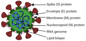
Coronaviruses are a large family of enveloped RNA viruses capable of infecting humans and animals. Among them, severe acute respiratory syndrome coronavirus (SARS-CoV) and severe acute respiratory syndrome coronavirus 2 (SARS-CoV-2) are highly pathogenic viruses associated with severe respiratory diseases. SARS-CoV-2, first identified in Wuhan, China in late 2019, rapidly spread worldwide and became the causative agent of Coronavirus Disease 2019 (COVID-19). The infection can progress from mild respiratory symptoms to severe pneumonia, acute respiratory distress syndrome (ARDS), multi-organ failure, and death.
A major factor responsible for the infectivity and pathogenicity of SARS-CoV-2 is the spike glycoprotein, commonly known as the spike protein or S protein. This viral surface protein is essential for host cell recognition, viral attachment, membrane fusion, and viral entry into human cells. Because of its critical biological role and strong immunogenicity, the spike protein has become the principal target for COVID-19 vaccines, monoclonal antibody therapies, and antiviral drug development.
Structure and Biological Organization of the SARS-CoV-2 Spike Protein
The SARS-CoV-2 spike protein is a transmembrane homotrimeric class I fusion glycoprotein located on the viral envelope. Each spike monomer has an approximate molecular weight of 180 kDa and forms a crown-like structure on the viral surface, giving coronaviruses their characteristic appearance under electron microscopy.
The spike protein exists in a metastable prefusion conformation and is composed of two major functional subunits:
- S1 subunit: responsible for host receptor recognition and attachment.
- S2 subunit: responsible for fusion between viral and host cell membranes.
The S1 region contains the receptor-binding domain (RBD), which specifically recognizes the human angiotensin-converting enzyme 2 (ACE2) receptor. ACE2 is highly expressed on epithelial cells of the respiratory tract, particularly in type II pneumocytes of the lungs. Binding of the RBD to ACE2 is the first critical step in SARS-CoV-2 infection.
The S2 subunit contains the viral fusion machinery, including heptad repeat domains, fusion peptides, and transmembrane regions required for membrane fusion and viral entry.
Mechanism of SARS-CoV-2 Cell Entry
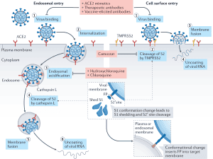
Viral entry requires coordinated interactions between the spike protein and host proteases. After attachment of the RBD to ACE2, the spike protein undergoes proteolytic activation by host cell enzymes, particularly transmembrane protease serine 2 (TMPRSS2).
TMPRSS2 cleaves and primes the spike protein at specific cleavage sites, enabling structural rearrangements necessary for membrane fusion. This process allows the viral envelope to fuse with the host cell membrane, facilitating viral genome release into the cytoplasm.
One unique structural characteristic of SARS-CoV-2 is the presence of a polybasic furin cleavage site at the S1/S2 junction. This cleavage sequence contains multiple arginine residues and is absent in the original SARS-CoV spike protein.
The furin cleavage region can be represented as:
RRARRRARRRAR
Furin is a ubiquitously expressed host protease found in many human tissues. The presence of this cleavage site increases viral activation efficiency and may contribute to:
- enhanced viral infectivity,
- broader tissue tropism,
- increased transmissibility,
- and higher pathogenic potential.
This structural adaptation is considered one of the important molecular features distinguishing SARS-CoV-2 from several related coronaviruses.
Receptor-Binding Domain (RBD) and ACE2 Recognition
The receptor-binding domain is one of the most important structural elements of the SARS-CoV-2 spike protein. Located at the apex of the S1 subunit, the RBD mediates direct interaction with ACE2 receptors.
The RBD undergoes dynamic conformational changes between two functional states:
- Up (open) conformation: receptor-accessible state allowing ACE2 binding.
- Down (closed) conformation: receptor-hidden state reducing immune recognition.
This conformational flexibility helps the virus balance efficient host cell attachment with partial immune evasion.
The ACE2 interaction mechanism involves multiple amino acid residues within the receptor-binding motif (RBM), a specialized region inside the RBD. Structural studies using cryogenic electron microscopy (cryo-EM) and X-ray crystallography demonstrated that SARS-CoV-2 possesses a larger binding interface and stronger affinity for ACE2 compared with SARS-CoV.
The receptor interaction can be summarized as:
SARS-CoV-2 RBD+ACE2→Virus Attachment and Entry
This higher receptor affinity partially explains the elevated transmissibility and infectivity of SARS-CoV-2.
Glycosylation and Immune Evasion of the Spike Protein
The spike protein is heavily glycosylated and contains numerous N-linked glycan sites distributed across both S1 and S2 subunits.
Each monomer contains approximately:
- 13 glycans in the S1 region,
- 9 glycans in the S2 region.
In the complete spike trimer, this corresponds to nearly 66 glycosylation sites.
These glycans perform several important biological functions:
- stabilization of protein folding,
- regulation of protease processing,
- protection against enzymatic degradation,
- shielding of viral epitopes from antibody recognition,
- modulation of immune responses.
Although SARS-CoV-2 is glycosylated, its spike protein is less densely shielded compared with certain other viral glycoproteins such as HIV-1 envelope proteins. Reduced glycan density may improve immune accessibility and contribute to stronger humoral immune responses.
Both N-linked and O-linked glycans have been identified on the spike surface, especially around the RBD. These carbohydrate structures may interfere with antibody accessibility and influence vaccine efficacy.
Structural Analysis Using Cryo-EM and X-Ray Crystallography
Advanced structural biology techniques have provided detailed insights into spike protein architecture.
High-resolution cryo-electron microscopy revealed that the spike protein forms a trimeric prefusion structure with a triangular cross-section. Structural investigations also identified the dynamic behavior of the RBD during receptor engagement.
X-ray crystallography studies further characterized the molecular interaction between the SARS-CoV-2 RBD and ACE2 receptor at near-atomic resolution. The RBD core contains a twisted antiparallel β-sheet structure linked with helices and loops forming the receptor-binding motif.
The receptor-binding affinity difference between SARS-CoV and SARS-CoV-2 can be represented by equilibrium dissociation constants:
KD(SARS-CoV-2)≈4.7 nM<KD(SARS-CoV)≈31 nM
This stronger ACE2 interaction significantly contributes to viral spread and efficient infection.
Humoral Immune Response Against the Spike Protein
The spike protein is highly immunogenic and serves as the major target of neutralizing antibodies generated during SARS-CoV-2 infection.
Convalescent COVID-19 patients produce robust adaptive immune responses involving:
- CD4+ T lymphocytes,
- CD8+ T lymphocytes,
- neutralizing IgG antibodies,
- IgM antibodies,
- IgA antibodies.
Most neutralizing antibodies target the receptor-binding domain because blocking ACE2 interaction directly inhibits viral entry.
Several potent monoclonal antibodies have demonstrated strong neutralizing activity against SARS-CoV-2, including:
- BD-368-2,
- B38,
- H4,
- CR3022,
- S309,
- 47D11.
Some antibodies recognize conserved epitopes shared between SARS-CoV and SARS-CoV-2, enabling partial cross-neutralization. Others are highly specific to SARS-CoV-2 RBD structures.
Research also demonstrated that camelid-derived nanobodies targeting spike RBDs can efficiently neutralize SARS-CoV-2 pseudoviruses. These findings highlight the importance of spike-focused immunotherapy and antibody engineering.
Vaccine Development Targeting the Spike Protein
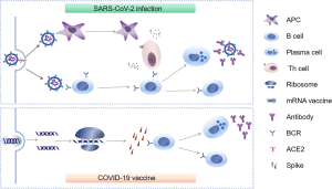
Due to its surface exposure and strong antigenicity, the spike protein rapidly became the primary antigen used in COVID-19 vaccine development.
Multiple vaccine technologies were developed using either the full-length spike protein or the receptor-binding domain:
- mRNA vaccines,
- DNA vaccines,
- adenoviral vector vaccines,
- recombinant protein vaccines,
- nanoparticle vaccines.
A major advancement in vaccine engineering involved stabilizing the spike protein in its prefusion state. Stabilized variants preserve important neutralizing epitopes and improve immune recognition.
One important stabilized spike design is the HexaPro variant, which exhibits enhanced structural stability and increased antigenicity.
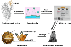
Experimental vaccine studies in rhesus macaques, mice, and guinea pigs demonstrated:
- strong neutralizing antibody production,
- activation of CD4+ and CD8+ T cell responses,
- reduced viral load after viral challenge,
- protection against respiratory infection.
DNA vaccine candidates such as INO-4800 and adenoviral vector vaccines such as ChAdOx1 nCoV-19 showed promising immunogenicity and protective efficacy in preclinical and clinical studies.
The overall vaccine strategy can be summarized as:
Spike Protein Antigen→Adaptive Immune Response→Neutralizing Antibodies and T Cell Immunity
Importance of Spike Protein Research in COVID-19 Control
Understanding the structural and functional characteristics of the SARS-CoV-2 spike protein remains essential for:
- vaccine optimization,
- antiviral drug discovery,
- monoclonal antibody development,
- diagnostic assay design,
- surveillance of emerging viral variants.
Mutations within the RBD and furin cleavage site can influence viral transmissibility, immune escape, and vaccine effectiveness. Therefore, continuous structural monitoring of spike protein evolution is critical for public health and pandemic preparedness.
Conclusion
The SARS-CoV-2 spike protein is the central molecular machinery responsible for viral attachment, membrane fusion, and host cell entry. Its complex structural organization, dynamic receptor-binding domain, glycosylation patterns, and interaction with ACE2 collectively determine viral infectivity and immune recognition.
Advanced structural biology studies using cryo-EM and crystallography have significantly improved understanding of spike protein architecture and receptor interactions. These discoveries accelerated the development of vaccines, monoclonal antibodies, and therapeutic interventions against COVID-19.
Because the spike protein is highly immunogenic and exposed on the viral surface, it remains the most important target for adaptive immunity and vaccine design. Continued investigation of spike protein structure, mutations, and immune interactions will remain essential for controlling SARS-CoV-2 infection and improving future coronavirus vaccine strategies.